The Zygomatic Arch Is Composed of Which of the Following
This definition incorporates text from the wikipedia website - Wikipedia. Lies posterior and inferior and joins with the zygomatic process of the temporal bone to form the zygomatic arch.

Illustration Of The Zygomatic Arch Masseter Muscle And Strain Gauge Download Scientific Diagram
Check all that apply ________ a.
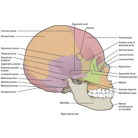
. Zygomatic arch bridge of bone extending from the temporal bone at the side of the head around to the maxilla upper jawbone in front and including the zygomatic cheek bone as a major portion. The zygomatic arch forms the ventral and lateral rim of the orbit. Note that the process is named not for the bone it is extending from but the bone.
Palatine process of maxilla. The Zygomatic Arch is a bridge of bone on the lateral head which is composed of zygomatic processes of the temporal and zygomatic bones. The zygomatic arch is composed of which of the following.
The incision is made deeply to the subaponeurotic areolar tissue following about 4 cm posterior to the hairline. Temporaral process of the zygomatic E. It then appears as if twisted inward upon itself and runs forward its surfaces now looking medialward.
Inserts on the angle and ramus of the mandible c. The malar surface of the zygomatic bone is perforated near its center which gives rise to the zygomaticofacial foramen. A 24-year-old female presented with complaint of right zygomatic arch pain following orthodontic treatment.
The medical history is. Its a foramen the zygomatico-orbital foramen which carries a zygomatic nerve. Medical Definition of zygomatic arch.
Frontal process of the zygomatic B. Due to its size and function in joining many facial bones together underdeveloped zygomatic bones cause significant issues related to the construction of the face. Palatine process of the maxilla.
The zygomatic arch is made up of the zygomatic bone and part of the. Frontal process of the zygomatic bone. The Zygomatic Arch plays a structural and protective role for muscles and nerves of.
The ________ attaches to the zygomatic arch and also to the angle of the mandible. A part of the lateral wall and floor of the Orbital surface. Temporal process of the zygomatic bone.
History of present illness. Slightly below this foramen is an elevation which serves as the origin to the zygomaticus. It presents 3 surfaces viz.
The cranial portion of the zygomatic arch is formed by the zygomatic bone and the caudal portion is formed by the zygomatic process of the temporal bone. Mandibular movements tended to aggravate the pain. The bony arch in vertebrates that extends along the side or front of the skull beneath the eye socket and that is formed by the zygomatic bone and the zygomatic process of the temporal bone.
Mental foramen of the mandible. All of the above statements are true 1 See answer haleygrobertson4125 is waiting for your help. This muscle originates on the zygomatic arch and inserts at the angle and ramus of the mandible.
The arch of bone that extends along the front or side of the skull beneath the orbit and that is formed by the union of the temporal. The zygomatic process of the temporal bone is a long arched process projecting from the lower part of the squamous portion of the temporal boneIt articulates with the zygomatic bone. This process is at first directed lateralward its two surfaces looking upward and downward.
Asked Sep 4 2019 in Anatomy Physiology by HoshGosh. The extension of the temporal bone is known specifically as the zygomatic process and attaches directly to the. Asked Sep 6 2019 in Anatomy Physiology by Redditor.
Up to 10 cash back The anatomy of the zygomatic bone is composed of two surfaces four processes and four borders. All of the following can occur EXCEPT a Biocompatibility tests conducted in vitro A primary infection of syphilis occurring on the tongue is referred to as aan One week following extraction of teeth 18 and 48 an 18 year old male returns to the dental office complaining of persistent bleeding from the extraction sites. Bony floor formed by the zygomatic frontal parietal sphenoid and the temporal bones attachment of the temporalis muscle.
The masseter muscle important in chewing arises from the lower edge of the arch. The patient stated that the pain had occurred 1 mo before the first visit and the pain level was 5 on a 0-10 NRS. The zygomatic bone is composed of the following 3 parts.
Another major chewing muscle the temporalis passes through the arch. Zygomatic arch -composed of the process temporal process of zygomatic extending from the jugal or zygomatic bone and a process extending from the temporal zygomatic process of temporal. It is responsible for closing the jaw.
Add your answer and earn points. Check all that apply A. Mental foramen of the mandible C.
The zygomatic arch is formed from parts of both the zygomatic bone and the temporal bone. 1 The arch forms the inferior boundary of the temporal fossa within which lies the temporalis muscle of mastication not pictured. Asked Jul 27 2018 in Anatomy Physiology by Jen66.
Is innervated by the trigeminal V nerve d. The zygomatic arch is composed of which of the following. Find the following parts of the human skull.
Orbital surface lateral surface and temporal surface. The frontal branch of the facial nerve lies under the superficial temporal fascia and where it crosses the zygomatic arch is separated only by the superficial fold of the temporal fascia and by areolar tissue. The zygomatic arch cheek bone is formed by the zygomatic process of temporal bone and the temporal process of the zygomatic bone the two being united by an oblique suture zygomaticotemporal suture.
The American Heritage Medical Dictionary Copyright 2007 2004 by Houghton Mifflin Company. The sagittal suture joins the two parietal bones. Originates on the zygomatic arch and the maxilla b.
The zygomatic bone consists of cartilage when a fetus is in utero with bone-forming immediately after birth. The bone that is the posterior component of the zygomatic arch is the. Zygomatic process of the temporal bone.
Palatine process of the maxilla D. The zygomatic arch cheek bone or zygoma are all interchangeable terms for the structure in the skull seen indicated by the arrow in the following image.
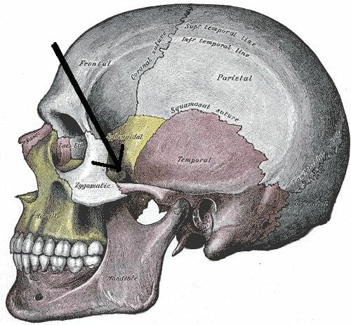
Zygomatic Arch Zygoma Cheek Bone Definition Quiz Biology Dictionary

Zygomatic Arch Radiology Reference Article Radiopaedia Org

The Relationship Between Morphometric Measurments Severity And Success Of Zygomatic Arch Fracture Reduction Journal Of Oral And Maxillofacial Surgery

Zygomatic Arch Arcus Zygomaticus Image Yousun Koh Anatomie Schadel Anatomie Knochen

Temporomandibular Joint Temporomandibular Joint Facial Nerve Arteries And Veins
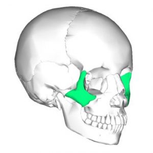
Ce4rt X Ray Positioning Guide For Radiologic Techs Zygomatic Arch

Last Week S Mysteryanatomy Structure Was The Zygomatic Arch The Zygomatic Arch Is Form By The Articulation Of Biology Teacher Science Student Life Science
Is The Cheekbone In A Rabbit Called The Maxilla Or The Zygomatic Bone Somewhere It Is Mentioned That It Is The Maxilla And Somewhere Else It Is The Zygomatic Bone In My

Illustration Of The Zygomatic Arch Masseter Muscle And Strain Gauge Download Scientific Diagram
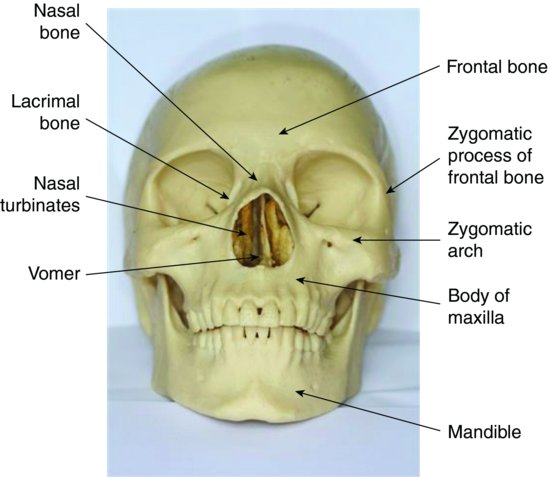
7 Skull And Oral Anatomy Pocket Dentistry

The Skull Bone Anatomy And Physiology By Er Services Skull Anatomy Anatomy And Physiology Human Anatomy And Physiology

Close Up Lateral View Skull Netter Anatomy Bones Anatomy Nursing School Notes

Lateral Aspect Of Orbit Zygomatic Arch Ramus Of Mandible And Download Scientific Diagram

Chapter 8 The Skeletal System Ppt Download

Exercise 7 Facial Bones Thoracic Cage Thoracic Vertebrae

Pin On Paleoanthropology Paleoantropologia
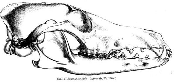
Zygomatic Arch Zygoma Cheek Bone Definition Quiz Biology Dictionary
:watermark(/images/watermark_only_sm.png,0,0,0):watermark(/images/logo_url_sm.png,-10,-10,0):format(jpeg)/images/anatomy_term/processus-zygomaticus-ossis-temporalis/qV8PrsGuLEtpPqAmybaeiA_861_Skull_Lateral_View.png)
Temporal Bone Anatomy Parts Sutures And Foramina Kenhub

Aneurysmal Bone Cyst Of The Zygomatic Arch A Case Report Clinical Imaging
Comments
Post a Comment